All Three Regions of the Brain Stem Can Be Observed on the Ventral Surface of the Brain.
Original Editor - Naomi O'Reilly
Top Contributors - Naomi O'Reilly, Lucinda hampton, Kim Jackson, Tarina van der Stockt, Admin, Simisola Ajeyalemi, Shaimaa Eldib, Olajumoke Ogunleye, Aminat Abolade, Rachael Lowe, Tony Lowe and Wendy Walker
Introduction [edit | edit source]
The encephalon, contained in and protected past the skull and suspended in cerebrospinal fluid, is one of the almost important and circuitous organs in the body. It is the key organ of the nervous system, and with the spinal cord makes upward the central nervous system, which controls nigh of the activities of the body, processing, integrating, and analogous the information information technology receives from the sense organs and determining the signals or instructions sent back to the rest of the body. [1]
At birth, the average brain weighs near 350 - 400grams, approximately 25% of the final adult encephalon weight of i.four - 1.45 kg and accounting for only 2% of overall body mass, which is reached between 10 and 15 years of age. Fastest growth occurs during the commencement 3 years of life, with virtually xc% of the adult value reached by the age of five years. Its average width is about 140 mm, the average length is about 167 mm, and average height virtually 93 mm. While the brain continues to modify throughout our life span, changes in brain morphology during babyhood, adolescence and adulthood are much more than subtle than those in the beginning four years of life.[ii] [3] [4] The rate and corporeality of growth that occurs in the brain later nascence is neither constant nor pre-determined, nor is it protected from outside influences, both positive and negative, and as such can be speeded up and increased, or slowed downward and decreased. [3]
Gross Beefcake [edit | edit source]
The brain consists of three main structural divisions; the cerebrum (outer layer is the cerebral cortex), the cerebellum, and the brain stem at the base of the brain, which extends from the upper cervical spinal cord to the diencephalon of the cerebrum. [v] [6]
Cerebrum [edit | edit source]
The cerebrum is the largest part of the brain. The surface of the cerebrum is composed of depressions or grooves (sulci) and ridges or raised areas (gyri), which increase the surface expanse of the cerebrum without an increase in the size of the brain. Grey thing, approximately 2 to 4 mm thick, forms the outer surface of the cerebrum, which processes and integrates data from white affair fibre tracts, which class the inner surface of the cerebrum. [7] The cerebrum consists of two cerebral hemispheres, the right hemisphere and the left hemisphere, continued past the corpus callosum which facilitates communication between both sides of the brain, with each hemisphere in the main connexion to the contralateral side of the body i.due east. the left hemisphere of the cerebrum receives data from the right side of the torso resulting in motor control of the correct side of the body and vice versa. The Hemispheres are then further divided into 4 lobes; [1] [7] [v]
Frontal Lobe [edit | edit source]
The frontal lobe is located at the front end of the brain, occupying the area anterior to the cardinal sulcus and superior to the lateral sulcus. It is associated with reasoning, motor skills, higher-level knowledge and expressive language. At the back of the frontal lobe, near the central sulcus, lies the motor cortex. This area of the encephalon receives information from various lobes of the brain and utilises this data to carry out body movements. Impairment to the frontal lobe tin lead to changes in sexual habits, socialisation, and attending besides as increased run a risk-taking. [1] [5] [6]
| Area | Part | Dysfunction |
|---|---|---|
| Principal Motor Cortex | Voluntary Control of Move | Altered Musculus Tone Poor Motor Command |
| Pre Motor Area | Selection of movement based on external events | Dyspraxia |
| Supplementary Motor Area | Pick of motility based on stored plans specified past internal cues. Involved in planning of motor actions. | |
| Pre-Supplementary Motor Surface area | Acquiring new sequences | Dyspraxia |
| Broca's Area | Left Hemisphere - Expression of Oral communication | Expressive Dysphasia |
| Correct Hemisphere - Non-Exact Advice | ||
| Pre-frontal Cortex | Personality and Behaviour | Changes in Grapheme |
| Inappropriate Behaviour | ||
| Higher executive function - problem solving, initiation, moderation and termination of bevaiour. | Dysexecutive Syndrome |
Parietal Lobe [edit | edit source]
The parietal lobe is located in the centre section of the brain, occupying an surface area posterior to the key sulcus and superior to the lateral sulcus, extending posteriorly as far as the parieto-occipital sulcus. Information technology is associated with processing tactile sensory information such as pressure, touch, and pain. A portion of the brain known as the somatosensory cortex is located in this lobe and is essential to the processing of the body'southward senses. [one] [5] [6]
| Area | Function | Dysfunction |
|---|---|---|
| Primary Somatosensory Cortex | Receives Sensory Data from Whole Body | Altered Sensation |
| Homunculus Representation | ||
| Sensory Clan Area | Integrates Sensory Information based on sensory inputs | |
| Left Hemisphere - Attention to Correct Side Simply | Inability to Perceive and Attend to Objects, Infinite or Own Body despite vision, somatosensation and motor ability being intact. |
| Correct Hemisphere - Attention to both Left and Right | ||
| Contralateral Neglect | ||
| Processing of Visual Data for perception of motor and spatial relationships | Visuospatial Dysfnction |
Temporal Lobe [edit | edit source]
The temporal lobe is located on the bottom section of the brain, occupying the expanse junior to the lateral sulcus. This lobe is also the location of the primary auditory cortex, which is important for interpreting sounds and the linguistic communication nosotros hear. The hippocampus is too located in the temporal lobe, which is why this portion of the brain is also heavily associated with the formation of memories. Impairment to the temporal lobe tin atomic number 82 to problems with memory, speech communication perception, and linguistic communication skills. [1] [v] [half-dozen]
| Area | Function | Dysfunction |
|---|---|---|
| Primary Auditory Cortex | Loudness | Deafness |
| Pitch | ||
| Localisation of Basic Sound | ||
| Temporal Association Areas: | Recognition and Identification fo Stimuli | Agnosia - Acknowledge the existence of a stimulus but unable to recognize what it is |
| Confront and Object Recognition | Prosopagnosia |
| Processing Complex Sound | |
| Recognize Spoken Word | Receptive Dysphasia |
| Interpret the pregnant of Speech | ||
| Process Visual Information related to class recognition and object representation | Visuoperceptual Dysfuncton |
| Linked to the storage of Long Term Memory | ||
| Pyriform Cortex | Processing Olfactory Sensations | Olfactory Dysfunction |
| Insula Cortex | Procedure Taste Awareness | Taste Dysfunction |
Occipital Lobe [edit | edit source]
The occipital lobe is located at the back portion of the brain, occupying the small area behind the parietal-occipital sulcus. It is associated with interpreting visual stimuli and information. The primary visual cortex, which receives and interprets information from the retinas of the eyes, is located in the occipital lobe. Damage to this lobe tin cause visual problems such every bit difficulty recognizing objects, an disability to identify colors, and trouble recognizing words. [1] [5] [half dozen]
| Expanse | Role | Dysfunction |
|---|---|---|
| Main Visual Cortex | Determines bones attributes of Vision
| Diplopia (Double Vision) |
| Blindness | ||
| Visual Clan Expanse | Reply to Visual Stimuli within Receptive Fields Modulated by Attention & Working Memory |
Cerebellum [edit | edit source]
The cerebellum, sometimes referred to as the "Petty Brain", is found inferior to the tentorium cerebella or tentorial membrane in the posterior cranial fossa, posterior to the quaternary ventricle, the pons and the medulla oblongata. The cerebellum accounts for approximately x % of the brain'due south total size but has for more than l % of the total number of neurones located in the entire brain. It is the largest function of the hindbrain forming the Deep Cerebellar Nuclei, which consists of an outer layer cortical region with an inner subcortical mass of cells. It consists of two cerebellar hemispheres joined past a narrow median vermis and is connected to the posterior aspect of the brainstem by iii cerebellar peduncles, which are symmetrical bundles of nervus fibres. The cerebellum is divided into iii minor lobes; anterior, middle and flocculonodular lobes, which receive information from the balance system of the inner ear, sensory fretfulness, and the auditory and visual systems. [ane] [6]
The cerebellum is primarily involved in the coordination of movements also as the learning of movements (motor learning) and has ipsilateral control of movement i.e. the left hemisphere of the cerebellum controls the left side of the body and vice versa. Information technology regulates initiation, timing, sequencing, and force generation of muscle contractions, sequencing the social club of muscle firing when a group of muscles work together to perform a movement and assists with balance and posture maintenance. While the cerebellum is associated with motor move and control, motor commands do not originate here, rather the cerebellum serves to modify these motor commands it receives from sensory systems of the spinal cord and from other parts of the brain, and integrates these inputs to fine-tune motor action in order to make motor movements authentic and useful. The cerebellum compares the intended movement originating from the motor cortex areas with the actual movement relayed back by the afferent systems and interneurons in the spinal string. The main functions of the cerebellum are to; [1] [5]
To deed as a comparator: comparison descending supraspinal signals with ascending afferent feedback. If there is any discrepancy, this is then fine-tuned to produce the actual movement desired via descending pathways. This helps to achieve smoothness and accuracy in movement.
To act as a timing device: information technology converts descending motor signals into a sequence of motor activation. This helps the movement achieve smoothness and coordination, maintaining posture and remainder. (receiving input from the vestibular system).
To initiate and store motion: has the ability to shop and update motor data. at that place is a significant role played inaccurate learned move. This is due to a modifiable synapse at the purkinje prison cell.
Cerebellar damage produces disorders in fine motion, equilibrium, posture, and motor learning depending on the part of the cerebellum involved and how information technology is damaged. Dysfunction may result in; Hypotonia (reduced musculus tone), Clutter, Dysarthria (the disability to articulate words properly), Nystagmus (rapid hasty eye movements) and Palatal Tremor / Myoclunus (tremor of the palate) [v] [6]
Brainstem [edit | edit source]
The brainstem evolutionarily is the most aboriginal role of the encephalon beginning at the foramen magnum occupying the posterior cranial fossa of the skull and is divided into iii regions; the medulla oblongata, the pons and the midbrain. It connects the narrow spinal cord with the forebrain. [i] [5] [six]
Medulla Oblongata [edit | edit source]
Located direct above the spinal cord at the level of the foramen magnum forming the base of the brain stem, and connecting the pons superiorly to the spinal cord inferiorly. The medulla oblongata is oblong in shape, approximately 3cm long with the descending tracts passing through it from two pyramids on the ventral surface of the medulla between the anterior median fissure, which is disrupted where the tracts cross the midline at the decussation of the pyramids. Cranial Nerves Ix (Glossopharyngeal), X (Vagus), XI (Accessory) and XII (Hypoglossal) emerge form the Medulla Oblongata. [1] [5] [6]
The Medulla Oblongata controls many vital autonomic functions such as middle rate, breathing, and blood pressure level, with the post-obit three master functions;
- to serve as a conduit for the ascending and descending tracts to connect the spinal cord to the higher centers in the forebrain,
- to contains reflex centres associated with command of respiration, the cardiovascular system and consciousness,
- to contains nuclei of Cranial Nerves Three and XII.
Pons [edit | edit source]
Located inductive to the cerebellum, the Pons connects the medulla oblongata to the midbrain. It is approximately ii.5cm long, composed of transverse fibres forming a ridge or bridge beyond the anterior surface area connecting the right and left cerebellar hemispheres, from which the pons gets its name. These fibres are the pontocerebellar fibers that are in plough projections from the corticopontine fibers. They cross to enter the contralateral heart cerebellar peduncle and thus enter the cerebellum. Cranial Nerves V (Trigeminal), Half-dozen (Abducens), VII (Facial) and 8 (Vestibulocochlear) emerge from the pons. [ane] [five] [6]
The Pons serves a number of important functions including playing a role in several autonomic functions including stimulation of breathing and decision-making slumber cycles.
Midbrain [edit | edit source]
Extending from the Pons to the Mamillary Body, the midbrain also termed the mesencephalon is the superior most attribute of the brainstem and considered the smallest region of the brain. The substantia nigra is closely associated with motor system pathways of the basal ganglia producing Dopamine which plays a role in the movement, movement planning, excitation, motivation. Cranial Nerves II (Optic), III (Oculomotor) and Iv (Trochlear) emerge form the midbrain. The midbrain is associated with vision, hearing, motor control, slumber/wake, arousal (alertness), and temperature regulation, acting as a sort of relay station for auditory and visual information. It contains the post-obit: [i] [5] [6]
- Nuclei for 10 of 12 Pairs of Cranial Fretfulness (All except the Olfactory and Optic Nerves)
- Apparatus for Decision-making Center Movements (Cranial Nerves Three, Four and VI )
- Monoaminergic Nuclei that project widely in the CNS
- Vital Respiration and Cardiovascular Centres
- Autonomic Centres
- Areas important for Consciousness
- Ascending and Descending Pathways, linking Encephalon to the Spinal Cord
Diencephalon [edit | edit source]
The diencephalon is located at the base of operations of the brain. It contains the: [1]
Thalamus [edit | edit source]
The thalamus, a large mass of greyness matter in the dorsal part of the diencephalon, serves as a relay station for ascending input to the cortex and receives information from each of the primal senses, except smell. It is hypothesised that the thalamus serves a gating function in filtering information. The thalamus as well plays an important role in regulating states of slumber and wakefulness [v] [6]
Epithalamus [edit | edit source]
The epithalamus, the posterior segment of the diencephalon, is made upwardly of the habenula, the habenular commissure, the posterior commissure, and the pineal gland. The function of the epithalamus is to connect the limbic system to other parts of the brain. Some functions of its components include the secretion of melatonin and secretion of hormones from the pituitary gland past the pineal gland circadian rhythms), and regulation of motor pathways and emotions. [5] [six]
Hypothalamus [edit | edit source]
The hypothalamus lies on either side of the 3rd ventricle, below the thalamus and between the optic chiasm and the midbrain. It receives a large input from limbic structures. It has a significantly big efferent output to the ANS and has a highly significant office in the control of pituitary endocrine function. [5] [half dozen]
The hypothalamus as well as inputting the ANS also has a large function to play in the homeostasis of many physiological systems such as hunger, thirst, water and sodium rest and temperature regulation. It too plays a role in retentiveness and emotional responses, providing autonomic and endocrine responses. It also plays a office in the control of circadian rhythms via retinal input to suprachiasmatic nucleus. At that place may also be hypothalamus input into sexual and emotional behaviour independent of its endocrine role. [v]
Limbic System [edit | edit source]
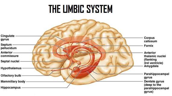
The limbic system is non defined past strict anatomic boundaries but incorporates several of import structures. The limbic structures conventionally include the amygdala, the hippocampus, the fornix, the mammillary bodies, the cingulate gyrus, and the parahippocampal gyrus, which lie mainly on the medial side of the temporal lobe. These structures grade connections between the limbic system and the hypothalamus, thalamus and cerebral cortex. [ane] [6]
The limbic organisation has been traditionally associated with our emotional behaviour. The hippocampus is important in retention and learning, while the limbic organisation itself is central in the control of emotional responses and provides loftier-level processing of sensory information. The master outflow of the limbic arrangement is to the prefrontal cortex and the hypothalamus as well as to cortical areas. Information technology appears to have a role in attaching behavioural significance and response to a given stimulus. Damage to this area has profound effects on emotional responses. [one] [5] [6]
While a full understanding of the limbic arrangement is far from consummate, advances in neurosciences have still given a meliorate understanding of the role the individual components of the limbic system play, and some insight into their many connections.[9]
Basal Ganglia [edit | edit source]
The basal ganglia are a group of large nuclei, which consist of the Neostratum (Caudate and Putamen), Internal and External segments of Globus Pallidus, the Pars Reticulata, the subthalamic nucleus and the compacta of Substantia Nigra, that partially environment the thalamus, which is important in the control of movement. The ruby nucleus and substantia nigra of the midbrain have connections with the basal ganglia.[half-dozen]
The basal ganglia are closely integrated with the motor cortex, premotor cortex, and motor nuclei of the thalamus and play a crucial function in modulation of movements. The Neostratum is the primary area of indicate reception for the basal ganglia and receives information from the whole cortex in a somatotopic fashion. Information technology relays signals to the thalamus, which projects to the premotor cortex, supplementary motor cortex and prefrontal cortex. There is as well a projection to the brainstem (pedunclopontine nucleus- involved in locomotion, and superior colliculus - eye movement). [1] [5]
Cranial Nerves [edit | edit source]
There are 12 pairs of cranial fretfulness, numbered according to the position where they originate in the inferior surface of the brain, that role mainly to convey motor signals to and sensory data from the head and cervix. The lower cranial nerves have somewhat more than complex visceral functions that are not strictly express to the head and cervix. The names of the Cranial Nerves (CN) are: [1] [5] [half dozen]
- CN I Olfactory
- CN II Optic
- CN Iii Oculomotor
- CN Four Trochlear
- CN V Trigeminal
- CN VI Abducens
- CN Seven Facial
- CN VIII Vestibulocochlear
- CN Nine Glossopharyngeal
- CN X Vagus
- CN XI Accompaniment
- CN XII Hypoglossal
Ventricles and Cerebrospinal Fluid [edit | edit source]
The ventricles are a arrangement of cavities/space within the cerebral hemispheres, which produce and broadcast cerebrospinal fluid, which is continuously produced and absorbed. The cerebrospinal fluid is produced, at a rate of about 450 mL/day, by the choroid plexuses within the lateral ventricles, and circulates through the ventricular organisation ultimately entering the subarachnoid infinite, which surrounds the cerebrum, cerebellum, brain stalk and spinal cord down to the level of the second sacral vertebrae. At whatsoever given time there is approximately 150 mL of cerebrospinal fluid, thus, the volume of CSF in most adults is turned over near iii times per twenty-four hour period. [1] [6]
The encephalon has 4 ventricles, which is a series of interconnected spaces in the core of the encephalon. The lateral ventricles are within the cerebral hemispheres, which are connected to each other and to the third ventricle through a pathway chosen the Interventricular Foramen of Monro. The third ventricle lies in the midline, separating deeper brain structures such as the left and correct thalami. The third ventricle communicates with the fourth ventricle through the Cerebral Aqueduct of Sylvius, which is a long narrow tube. [1] [5]
Blood Supply [edit | edit source]
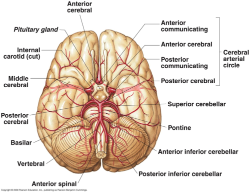
An adequate supply of oxygen and nutrients is vital for the normal part of the brain, which is achieved through a dense network of blood vessels. Arterial blood supply to the brain comes from 4 vessels the right and left Internal Carotid and the right and left Vertebral Arteries, which join at the base of the brain to grade the basilar artery. The Inductive Circulation is composed of the carotid arteries and their branches, while the Posterior Circulation is composed of the Vertobrobasilar arteries. [1] [v] [half-dozen]
| [x] | [xi] |
Internal Carotid Arteries [edit | edit source]
Traverse the skull within the carotid canal and clangorous sinus. They then pierce the dura and enter the middle cranial fossa lateral to the optic chiasm. They so divide to supply the anterior and heart sections of the cerebral hemispheres, anterior and center cerebral arteries. The internal carotid artery on each side terminates into the anterior cerebral artery, the middle cerebral artery, and the posterior communicating artery.[5]
Vertebral Arteries [edit | edit source]
Arising from the sub-clavian artery these ii arteries enter the skull through foramen magnum later passing through foramina in the transverse processes of the upper cervical vertebra. They unite in the brainstem to class the basilar avenue which ascends to form the two posterior cerebral arteries at the superior border of the Pons. This is the posterior cerebral hemisphere blood supply. This network is termed the posterior circulation. On its journey to becoming the basilar avenue, the vertebral arteries give off a number of branches, including the posterior spinal artery, posterior inferior cerebral artery and the anterior spinal artery. These constitute the blood supply to the upper cervical cord. The posterior junior cerebral artery supplies the lateral medulla and cerebellum. Impairment to these blood vessels can consequence in motor loss or sensory loss. [5] [6]
Basilar Artery [edit | edit source]
Formed by the ii vertebral arteries. This has a number of branches: anterior and inferior cerebellar artery, artery to the labyrinth, pontine branches and superior cerebellar artery which supply the brain stalk and cerebellum. [5]
Inductive Cerebral Arteries [edit | edit source]
The anterior cerebral artery arises from the internal carotid, at the medial extremity of the lateral cerebral fissure. It passes frontwards and medialward across the anterior perforated substance, above the optic nerve, to the commencement of longitudinal crevice. Is one of a pair of arteries on the brain that supplies oxygenated blood to almost midline portions of the frontal lobes and superior medial parietal lobes. [5] [6]
Eye Cerebral Arteries [edit | edit source]
These supply parts of the frontal, temporal, occipital and parietal lobes bilaterally, with branches besides supplying the basal ganglia and posterior limb of the internal capsule.
Posterior Cerebral Arteries [edit | edit source]
These supply blood to the posterior parietal cortex, occipital lobe and inferior temporal lobe. There are several branches of this artery that supply the midbrain, thalamus, subthalamus, posterior internal capsule, optic radiations and cognitive peduncle.
Circle of Willis [edit | edit source]
The basilar avenue, the posterior cognitive arteries, the posterior communicating arteries, and the anterior cerebral arteries, along with the inductive advice artery, form an important collateral apportionment, the Anterior and Posterior Apportionment anastomose at the base of the brain, termed the Circle of Willis. These vessels lie inside the subarachnoid space and are a common location for cerebral aneurysms to form. [5] [6]
Venous Drainage [edit | edit source]
The venous apportionment of the brain is very different from that of the residual of the body. Usually arteries and veins run together as they supply and drain specific areas of the body, nevertheless, this is not the case in the brain. The major vein collectors are integrated into the dura to form venous sinuses, which are adjacent to the posterior cranial fossa. The venous sinuses collect the blood from the encephalon and pass information technology to the internal jugular veins. The superior and inferior sagittal sinuses bleed the cerebrum, the cavernous sinuses drains the anterior skull base. All sinuses eventually drain to the sigmoid sinuses, which exit the skull and form the jugular veins. These 2 jugular veins are essentially the only drainage of the brain. If occlusion of either of these venous systems and so raised intracranial pressure can develop. [1] [5] [6]
References [edit | edit source]
- ↑ i.00 1.01 1.02 ane.03 1.04 1.05 ane.06 i.07 i.08 one.09 1.x ane.xi i.12 1.xiii 1.xiv i.xv 1.sixteen 1.17 i.18 1.19 i.20 Moore KL, Agur AM, Dalley AF, Moore KL. Essential clinical anatomy. Wolters Kluwer Health,; 2015.
- ↑ Dekaban AS, Sadowsky D. Changes in brain weights during the span of human life: relation of brain weights to body heights and body weights. Annals of Neurology: Official Journal of the American Neurological Association and the Child Neurology Gild. 1978 October;4(four):345-56.
- ↑ 3.0 3.i Holmes GL, Milh MM, Dulac O. Maturation of the human brain and epilepsy. InHandbook of clinical neurology 2012 January 1 (Vol. 107, pp. 135-143). Elsevier.
- ↑ Hartmann P, Ramseier A, Gudat F, Mihatsch MJ, Polasek W. Normal weight of the brain in adults in relation to age, sex, body height and weight. Der Pathologe. 1994 Jun;15(iii):165-70.
- ↑ 5.00 5.01 v.02 five.03 v.04 5.05 5.06 v.07 5.08 five.09 5.x 5.11 5.12 5.13 five.14 5.15 5.xvi five.17 5.18 v.19 5.20 5.21 five.22 5.23 5.24 5.25 5.26 Snell RS. Clinical neuroanatomy. Lippincott Williams & Wilkins; 2010.
- ↑ half dozen.00 6.01 6.02 6.03 six.04 six.05 half-dozen.06 6.07 vi.08 vi.09 vi.10 6.11 six.12 six.13 vi.14 6.15 6.16 6.17 6.xviii 6.19 6.twenty six.21 half dozen.22 half dozen.23 Jones, 1000. Neurological Assessment A Clinicians Guide. Churchill Livingstone Elseiver, 2011.
- ↑ 7.0 7.1 FitzGerald MJT, Gruener G, Mtui E: Clinical neuroanatomy and neuroscience, St Louis, 2012, Elsevier, pp 78, 97–110, 299.
- ↑ UBC Medicine - Educational Media. The Cerebellum - UBC Neuroanatomy - Season 1 - Ep 8. Available from: https://youtu.exist/17mxfO9nklQ[last accessed xxx/08/19]
- ↑ Torrico TJ, Abdijadid Southward. Neuroanatomy, Limbic System. InStatPearls [Internet] 2022 February 10. StatPearls Publishing.Available from: https://www.ncbi.nlm.nih.gov/books/NBK538491/ (last accessed xi.10.2019)
- ↑ khanacademymedicine. Cerebral blood supply - Part one. Bachelor from: http://www.youtube.com/watch?v=hfG8J_X1D5Q [last accessed 29/08/sixteen]
- ↑ khanacademymedicine. Cerebral blood supply - Role ii. Available from: http://world wide web.youtube.com/lookout man?v=kVulo3qDcUo [last accessed 29/08/16]
- ↑ AnatomyZone. Circumvolve of Willis - 3D Anatomy Tutorial. Bachelor from: http://world wide web.youtube.com/watch?v=9hhfM7rQHiM[final accessed thirty/10/17]
Source: https://www.physio-pedia.com/Brain_Anatomy
0 Response to "All Three Regions of the Brain Stem Can Be Observed on the Ventral Surface of the Brain."
Post a Comment